Home » Without Label » Anatomy Of Chest : Normal Female Anatomy Of The Chest Thoracic Cavity And Lungs Medical Art Works - Anatomy of the chest and stomach, human anatomy, anatomy of the chest and stomach.
Anatomy Of Chest : Normal Female Anatomy Of The Chest Thoracic Cavity And Lungs Medical Art Works - Anatomy of the chest and stomach, human anatomy, anatomy of the chest and stomach.
Anatomy Of Chest : Normal Female Anatomy Of The Chest Thoracic Cavity And Lungs Medical Art Works - Anatomy of the chest and stomach, human anatomy, anatomy of the chest and stomach.. Anatomy of the thorax, heart, abdomen and pelvis recommended text gray's anatomy for students. The chest is the area of origin for many of the body's systems as it houses organs such as the heart, esophagus, trachea, lungs, and thoracic diaphragm. The muscles of the chest develop from the somites found in the mesoderm. Thoracic wall the first step in understanding thorax anatomy is to find out its boundaries. 30 lines of the thoracic wall syllabus p.
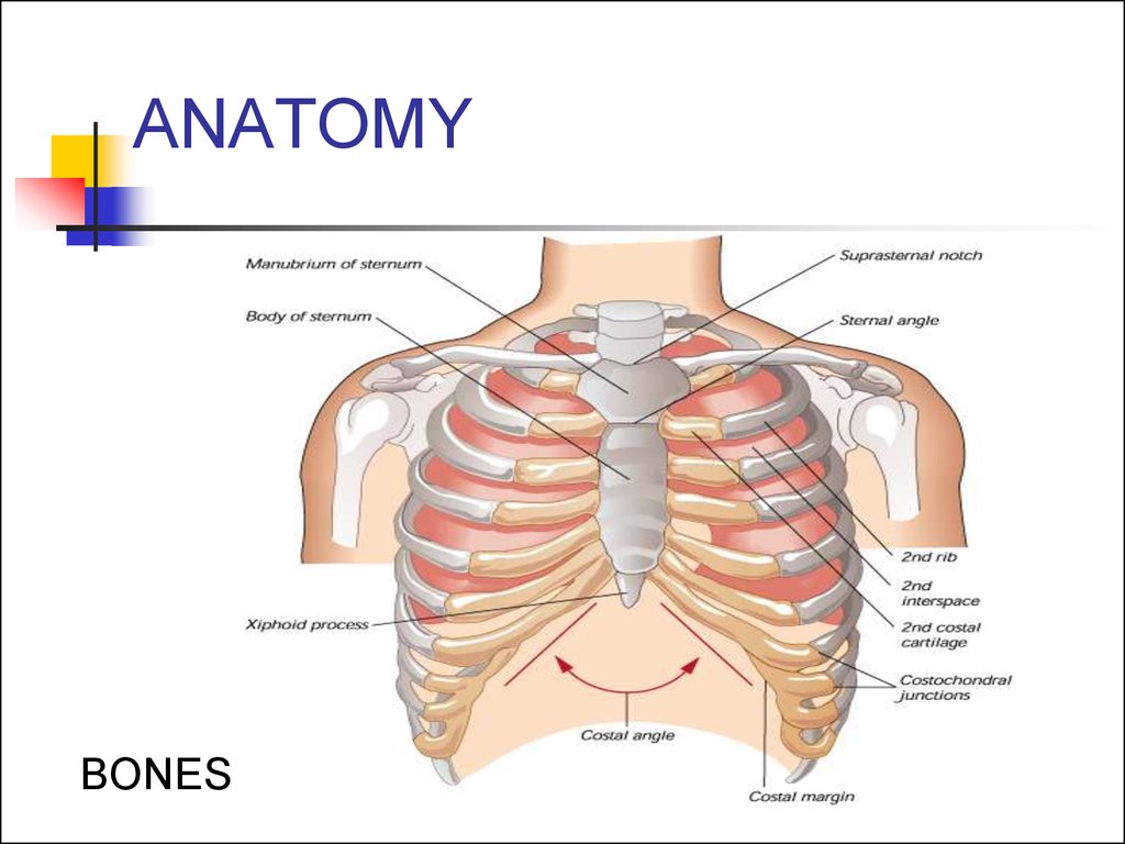
Thoracic wall the first step in understanding thorax anatomy is to find out its boundaries. A good radiologist knows the anatomy because knowing where structures normally live and recognizing the location of an abnormality helps to make or narrow the differential diagnosis. Thoracic cavity, also called chest cavity, the second largest hollow space of the body. Swensen fund for innovation in teaching. Learn about each of these muscles, their locations, functional anatomy and exercises for them.
Imaging Anatomy Chest Abdomen Pelvis Michael P Federle 9780323477819 from d1w7fb2mkkr3kw.cloudfront.net These myotomes divide into the epimere and the hypomere. Here, we break down the anatomy of your chest muscles. The myotomes elongate and invade the mesoderm of the wall of the embryonic thoracic and abdominal cavities. Anatomy of the thorax, heart, abdomen and pelvis recommended text gray's anatomy for students. Thoracic wall the first step in understanding thorax anatomy is to find out its boundaries. See chest anatomy stock video clips. Computed tomography (ct) of the chest can detect pathology that may not show up on a conventional chest radiograph(1). Swensen fund for innovation in teaching.
It provides protection to vital organs (eg, heart and major vessels, lungs, liver) and provides stability for movement.
Swensen fund for innovation in teaching. See chest anatomy stock video clips. See human chest anatomy stock video clips. Thoracic wall the first step in understanding thorax anatomy is to find out its boundaries. Three dimensional view of the female reproductive system, full frontal view. 31 anatomy of the female breast syllabus p. Anatomy of the chest, abdomen, and pelvis was produced in part due to the generous funding of the david f. 2 skin of the anterior chest wall syllabus p. The pectoralis major and the pectoralis minor, known collectively as your pecs. These myotomes divide into the epimere and the hypomere. Sternocleidomastoid muscle clavicle and ribs anatomy muscle anatomy chest sternocleidomastoid ribs anatomy chest muscles anatomy thorax rib muscles chest muscles chest anatomy illustration. 30 lines of the thoracic wall syllabus p. It is enclosed by the ribs, the vertebral column, and the sternum, or breastbone, and is separated from the abdominal cavity (the body's largest hollow space) by a muscular and membranous partition, the diaphragm.
Sternocleidomastoid muscle clavicle and ribs anatomy muscle anatomy chest sternocleidomastoid ribs anatomy chest muscles anatomy thorax rib muscles chest muscles chest anatomy illustration. The upper part of your pec major, the clavicular head runs from your clavicle (collarbone) across the top of your chest and attaches to your humerus, or upper arm. The pectoralis major and the pectoralis minor, known collectively as your pecs. The epidermis is the outermost layer that provides a protective, waterproof seal over the body. The circulatory system does most of its work.
Chest Pain Prezentaciya Onlajn from cf.ppt-online.org This atlas is a comprehensive and affordable learning tool for medical students and residents and especially for radiologists and pneumologists. Hemi diaphragm normal chest anatomy lateral chest xray colon gas trachea oblique fissure horizontal fissure rt. Anatomy of the thorax, heart, abdomen and pelvis recommended text gray's anatomy for students. Computed tomography (ct) of the chest can detect pathology that may not show up on a conventional chest radiograph(1). Anatomy of the female human body 12 photos of the anatomy of the female human body. About the 6th week, the somites differentiate into the sclerotomes and the dermatomyotomes. Use the mouse scroll wheel to move the images up and down alternatively use the tiny arrows (>>) on both side of the image to move the images.>>) on both side of the image to move the images. The epidermis is the outermost layer that provides a protective, waterproof seal over the body.
The chest is the area of origin for many of the body's systems as it houses organs such as the heart, esophagus, trachea, lungs, and thoracic diaphragm.
#anatomy of the chest and stomach. Related posts of anatomy of the chest area anatomy of the female human body. The chest or thorax is the region between the neck and diaphragm that encloses organs, such as the heart, lungs, esophagus, trachea, and thoracic diaphragm. It provides protection to vital organs (eg, heart and major vessels, lungs, liver) and provides stability for movement. Swensen fund for innovation in teaching. (1) the pectoralis major, and (2) the pectoralis minor. Anatomy of the thorax, heart, abdomen and pelvis recommended text gray's anatomy for students. Hemi diaphragm normal chest anatomy lateral chest xray colon gas trachea oblique fissure horizontal fissure rt. Radiology basics of chest ct anatomy with annotated coronal images and scrollable axial images to help medical students and junior doctors learning anatomy. Browse 6,407 chest anatomy stock photos and images available, or search for human anatomy to find more great stock photos and pictures. Principal functions are the protection of internal viscera and an expandable cylinder facilitating variable gas flow into the lungs. The circulatory system does most of its work. A good radiologist knows the anatomy because knowing where structures normally live and recognizing the location of an abnormality helps to make or narrow the differential diagnosis.
See human chest anatomy stock video clips. The chest anatomy includes the pectoralis major, pectoralis minor and the serratus anterior. Most people struggle to build the top portion of their chest, so we'll pay special attention to this area. Browse 6,407 chest anatomy stock photos and images available, or search for human anatomy to find more great stock photos and pictures. The muscles of the chest develop from the somites found in the mesoderm.
Chest Wall Anatomy Diagram Quizlet from o.quizlet.com Three dimensional view of the female reproductive system, full frontal view. Computed tomography (ct) of the chest can detect pathology that may not show up on a conventional chest radiograph(1). Plus, how to target each to make them bigger and stronger. This atlas is a comprehensive and affordable learning tool for medical students and residents and especially for radiologists and pneumologists. Browse 6,407 chest anatomy stock photos and images available, or search for human anatomy to find more great stock photos and pictures. See chest anatomy stock video clips. A good radiologist knows the anatomy because knowing where structures normally live and recognizing the location of an abnormality helps to make or narrow the differential diagnosis. 2 skin of the anterior chest wall syllabus p.
It spreads out like a fan and covers the rib cage like an armor plate.
Radiology basics of chest ct anatomy with annotated coronal images and scrollable axial images to help medical students and junior doctors learning anatomy. How to view the anatomical labels. The pectoralis major and the pectoralis minor, known collectively as your pecs. Here, we break down the anatomy of your chest muscles. Use the mouse scroll wheel to move the images up and down alternatively use the tiny arrows (>>) on both side of the image to move the images.>>) on both side of the image to move the images. The superior thoracic aperture found superiorly and the inferior thoracic aperture. Chest muscles anatomy (1) pectoralis major muscle. The chest wall is comprised of skin, fat, muscles, and the thoracic skeleton. These myotomes divide into the epimere and the hypomere. The upper part of your pec major, the clavicular head runs from your clavicle (collarbone) across the top of your chest and attaches to your humerus, or upper arm. Most people struggle to build the top portion of their chest, so we'll pay special attention to this area. Plus, how to target each to make them bigger and stronger. Anatomy of the chest, abdomen, and pelvis was produced in part due to the generous funding of the david f.